Jacobson et al. 2016: Coral nubbin IDs and experimental conditions
Project
Program
| Contributors | Affiliation | Role |
|---|---|---|
| Edmunds, Peter J. | California State University Northridge (CSUN) | Principal Investigator |
| Jacobson, Lianne | Contact | |
| York, Amber D. | Woods Hole Oceanographic Institution (WHOI BCO-DMO) | BCO-DMO Data Manager |
Methodology:
Sampling and analytical procedures:
We worked with small colonies of massive Porites spp., a functional group with unresolved taxonomy that is ecologically important and common on shallow reefs throughout the Indo-Pacific, and consists of P. lobata and P. lutea. We collected juvenile colonies (<40 mm diameter) of massive Porites spp. from the back reef (2–3 m depth) on the north shore of Moorea, French Polynesia. We collected corals twice, a few days prior to each trial, which began on 17 April 2010 (trial 1) and 8 May 2010 (trial 2).We chose massive Porites spp. for this study because: (i) members of this functional group have a thick tissue layer, and (ii) properties of coral tissue vary seasonally and are responsive to treatments including light, temperature and food supply. Therefore, we reasoned that starvation-induced changes in biomass would be relatively easy to detect and serve as a measure of treatment effects. Furthermore, the use of juvenile colonies (assumed to be <4 cm diameter, based on data for another species of the same genus) simplified the application of DEB theory (because allocation to reproduction could be ignored).
Following collection, we mounted each coral in a small disc (∼2 cm radius, 1 cm thick) of epoxy (Z-Spar A788, West Marine, Watsonville, CA, USA), molded to keep the coral nubbin upright and to fit snuggly into a custom-made respiration chamber. We allowed freshly prepared nubbins to acclimate to laboratory conditions (running seawater, pumped from the adjacent fringing reef) for ∼24 h prior to experimentation. Immediately after the first collection, we sampled 16 corals for initial estimates of respiration (N=8) and biomass (N=8), which served as a benchmark against which the effects of the treatments were compared. At the start of both trials, we recorded the buoyant weight of each coral (±1 mg).
Treatments: duration of starvation We conducted two consecutive trials of a similar experiment. The two trials increased replication, and thus the possibility of detecting the effects of starvation. Logistical constraints prevented us from conducting a single experiment with a larger number of replicate corals and multiple treatments of starvation duration. The two trials employed the same starvation conditions to elicit a metabolic response, but the results of the first trial motivated sampling with an increased temporal resolution in the second trial. Every 4 days during the first trial and every 2 days during the second trial, we determined the respiration, biomass and skeletal mass of 6 corals to evaluate the response to different durations of starvation (i.e. treatments). We conducted both trials in tanks containing ∼45 l of seawater; during trial 1, an acrylic aquarium (100 l) was used, and in trial 2, an insulated aquarium (50 l) was used. Pumps (ViaAqua VA-1380, 1380 l h−1, Commodity Axis Inc., Camarillo, CA, USA; and Rio 8HF Hyper Flow, 2079 l h−1, TAAM, Camarillo, CA, USA) circulated the enclosed seawater, to maintain the air saturation of the seawater (i.e. ∼21% O2 as assessed with a fiber optic O2 electrode; Foxy-R/Foxy-AF, Ocean Optics, Dunedin, FL, USA). We maintained the temperature of the seawater in the tanks at the mean ambient temperature of seawater in the back reef of Moorea in April when the experiment was completed (29.40±0.06°C, mean±s.e., N=39), and we measured seawater temperature 2–3 times daily using a certified thermometer (YSI series 400 probe, ±0.05°C accuracy, cat. no. 15-077-8, Fisher Scientific, Waltham, MA, USA).
To starve the corals of autotrophic and heterotrophic food resources, we covered the tanks in black plastic to exclude light, and filtered seawater (0.2 μm final pore size in fiberglass/ polypropylene pleated filters; Heyes Filters, Torrance, CA, USA) to exclude particulate food. The pore size of the filters was small enough to remove bacteria, and filtration was enhanced by first sterilizing the seawater (Current USA Gamma UV Sterilizer, 8 W T5 lamp; G. Heyes, personal communication). Furthermore, we reduced the concentration of dissolved organic materials (normally, total organic carbon is ∼37 mmol l−1 and particulate organic carbon is ∼3 mol l−1 in near-shore seawater in Moorea by passing the seawater through a column filled with granules of activated carbon (Boyd, Chemi Pure Elite). Once daily, we replaced ∼30% of the seawater within the tank with seawater freshly collected offshore and filtered as described above.
Response variables
We assessed the metabolic rate of corals by measuring dark respiration, which was recorded using an acrylic chamber (0.285 l), containing a magnetic stir bar to create water motion. Located marginally within the chamber, the coral received radial flow at ∼3.5±0.3 cm s−1 (mean±s.e., N=9), as estimated by photographing brine shrimp eggs. A water jacket and chiller (RE106 and E100, Lauda Brinkmann, Delran, NJ, USA) maintained the temperature (29.0°C) of the filtered seawater (0.2 μm) that filled the chamber. An oxygen probe (Foxy-R/Foxy- AF, Ocean Optics) that functioned with a spectrophotometer (USB2000, Ocean Optics) continually recorded the partial pressure of O2 in the chamber. We calibrated the probe using a two-point calibration with water-saturated air and a saturated solution of sodium sulfite (NaSO3) in 0.01 mol l−1 sodium tetraborate (Na2B4O7) as the 21% and 0% O2 saturation standards, respectively. We calculated O2 concentrations from O2 saturation using tabulated values for solubility of O2 as a function of temperature and salinity. We measured salinity with a conductivity meter (YSI 3100, Yellow Springs, OH, USA). Each coral was incubated in the respiration chamber for 15–60 min, or until O2 saturation decreased ≥5%, but never fell below 85% to avoid hypoxia, which could affect respiration. We quantified the respiration rate of a coral as the difference between the O2 depletion in a chamber containing a coral and filtered seawater and the rate of oxygen depletion in a chamber containing only filtered seawater. We estimated the surface area of coral tissue using aluminium foil, and then standardized respiration rates by that surface area (μmol O2 cm−2 day−1).
We measured biomass by preserving corals in 5% formalin for 24 h, then dissolving their skeletons in 10% hydrochloric acid over ∼24–48 h. Decalcification was completed outdoors because of the possible formation of carcinogenic gas when hydrochloric acid is mixed with formalin. We rinsed decalcified tissue tunics in freshwater and removed fouling organisms (e.g. algae) with forceps, then dried the tunics to a constant mass at 60°C. Dry mass was converted to carbon mass, assuming 0.442 mol C g−1 (see Appendix for details). Following different durations of starvation (i.e. the treatments), we again recorded the buoyant weight of the corals, and used the difference from the initial buoyant weight to calculate calcification. While recording the buoyant weight, we kept the seawater at ambient temperature (29°C) and suspended the corals from monofilament thread attached to a top-loading balance (PB153-S, ±1 mg, Mettler Toledo, Columbus, OH, USA). We converted the change in buoyant weight to a change in skeleton dry mass based on empirically determined seawater density and the density of aragonite (2.93 g cm−3), and skeletal mass was converted to mol CaCO3 cm−2 using the molecular mass of CaCO3.
BCO-DMO Data Manager Processing Notes:
* Original data submitted as in Excel sheet "Nubbin ID" extracted to csv. See Data Files for the originally submitted Excel file.
* added a conventional header with dataset name, PI name, version date
* modified parameter names to conform with BCO-DMO naming conventions (spaces, +, and - changed to underscores). Units in parentheses removed and added to Parameter Description metadata section.
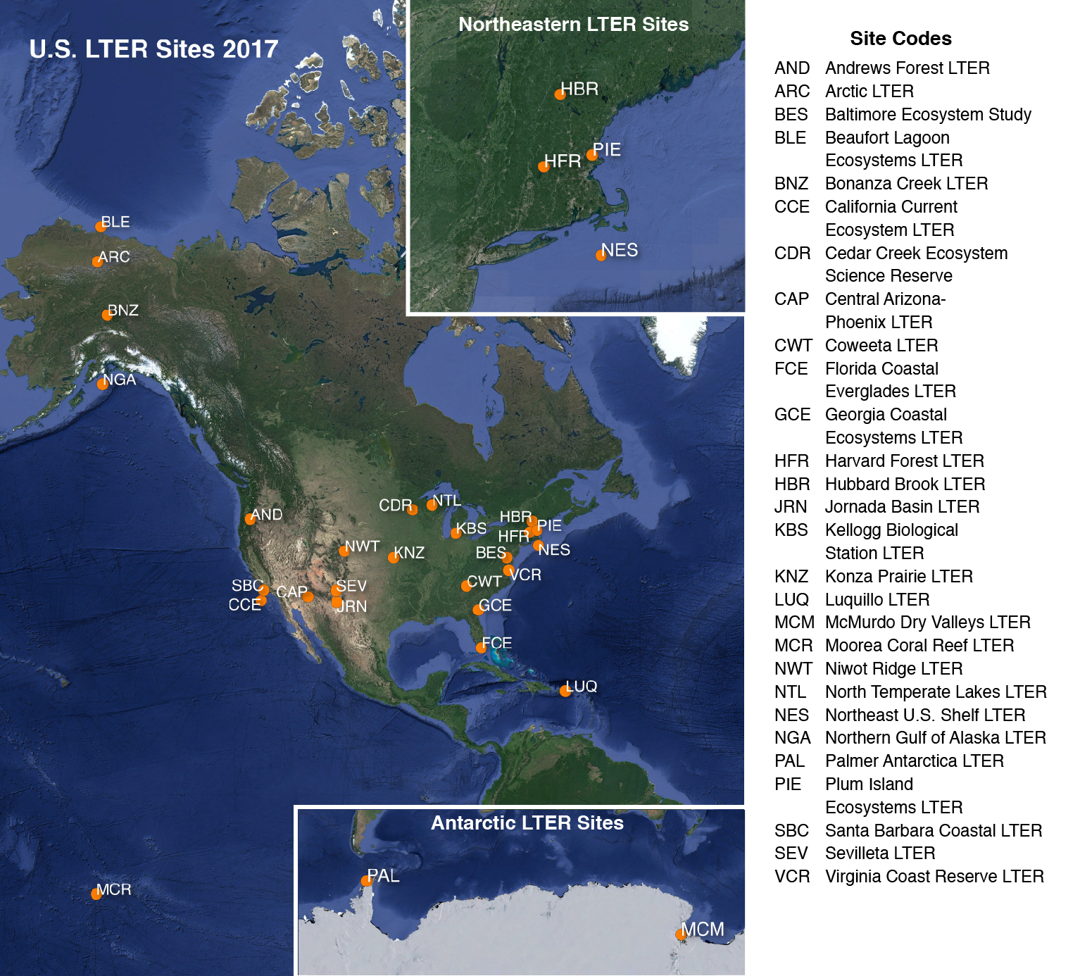
Moorea Coral Reef Long-Term Ecological Research site (MCR LTER)
From http://www.lternet.edu/sites/mcr/ and http://mcr.lternet.edu/:
The Moorea Coral Reef LTER site encompasses the coral reef complex that surrounds the island of Moorea, French Polynesia (17°30'S, 149°50'W). Moorea is a small, triangular volcanic island 20 km west of Tahiti in the Society Islands of French Polynesia. An offshore barrier reef forms a system of shallow (mean depth ~ 5-7 m), narrow (~0.8-1.5 km wide) lagoons around the 60 km perimeter of Moorea. All major coral reef types (e.g., fringing reef, lagoon patch reefs, back reef, barrier reef and fore reef) are present and accessible by small boat.
The MCR LTER was established in 2004 by the US National Science Foundation (NSF) and is a partnership between the University of California Santa Barbara and California State University, Northridge. MCR researchers include marine scientists from the UC Santa Barbara, CSU Northridge, UC Davis, UC Santa Cruz, UC San Diego, CSU San Marcos, Duke University and the University of Hawaii. Field operations are conducted from the UC Berkeley Richard B. Gump South Pacific Research Station on the island of Moorea, French Polynesia.
MCR LTER Data: The Moorea Coral Reef (MCR) LTER data are managed by and available directly from the MCR project data site URL shown above. The datasets listed below were collected at or near the MCR LTER sampling locations, and funded by NSF OCE as ancillary projects related to the MCR LTER core research themes.
This project is supported by continuing grants with slight name variations:
- LTER: Long-Term Dynamics of a Coral Reef Ecosystem
- LTER: MCR II - Long-Term Dynamics of a Coral Reef Ecosystem
- LTER: MCR IIB: Long-Term Dynamics of a Coral Reef Ecosystem
- LTER: MCR III: Long-Term Dynamics of a Coral Reef Ecosystem
- LTER: MCR IV: Long-Term Dynamics of a Coral Reef Ecosystem
Long Term Ecological Research network (LTER)
adapted from http://www.lternet.edu/
The National Science Foundation established the LTER program in 1980 to support research on long-term ecological phenomena in the United States. The Long Term Ecological Research (LTER) Network is a collaborative effort involving more than 1800 scientists and students investigating ecological processes over long temporal and broad spatial scales. The LTER Network promotes synthesis and comparative research across sites and ecosystems and among other related national and international research programs. The LTER research sites represent diverse ecosystems with emphasis on different research themes, and cross-site communication, network publications, and research-planning activities are coordinated through the LTER Network Office.
2017 LTER research site map obtained from https://lternet.edu/site/lter-network/
| Funding Source | Award |
|---|---|
| NSF Division of Ocean Sciences (NSF OCE) |
[ table of contents | back to top ]
